 Reichelt Chemietechnik Magazine
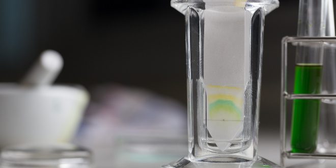
Where the history of chromatography begins remains uncertain. Nature itself performs the process: a chemical mixture separates into its individual components as it passes through an environment made of different materials. Around 1900, the American geochemist David Talbot Day (1859 – 1925) observed one such case. He found that the components of crude oil, as they seep through certain porous or fine-grained rocks such as limestone, can sort themselves into long-chain, cyclic and aromatic hydrocarbons. In doing so, they leave differently coloured bands in the rock.
Ferdinand Runge’s “Chemical Coats of Arms”
Separating mixtures of dyes still serves as a form of entertainment today. Dab a blot of ink onto a piece of blotting or filter paper. Then add a few drops of diluted alcohol to the same spot. The paper absorbs the liquid, and the various dyes that make up the ink migrate outwards from the centre in rings. The result is a set of imaginative patterns. Similar images can be produced from plant extracts, using petroleum spirit as a “developing fluid” on absorbent paper.
One person recognised the potential of such images early on: the German chemist Friedlieb Ferdinand Runge (1794 – 1867). He explained them as “bonds formed through chemical interaction”. In 1855, he described them in detail in his treatise “Der Bildungstrieb der Stoffe, veranschaulicht in selbständig gewachsenen Bildern” (“The Formative Drive of Substances, Illustrated in Independently Grown Images”).
Spot Tests and pH Measurement
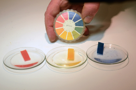
In a spot test, a drop of the dissolved sample is brought together with a specific reagent on a piece of filter paper. A change in colour then indicates the presence of a particular substance.
The familiar acid–base test using pH paper is one such spot test for pH measurement. The paper consists of filter-paper strips impregnated with an acid–base colour indicator. It fell to the Austrian chemist Fritz Feigl (1891 – 1971) to develop this simple spot test into a rapid analytical method. It remains in regular use in many chemical laboratories today.
Ring Chromatography and the Role of Capillary Action
Runge developed his “chemical coats of arms” on filter paper from mixtures of dyes, using various solvents. He watched how the solvents influenced the resulting colour patterns. He also let chemical reagents act on substances in the same way. By comparing these results with unknown substances, he could already draw conclusions about their composition. Runge’s “chemical coats of arms” can therefore be seen as a forerunner of ring chromatography. This variant of paper chromatography is now rarely used.
Among the first to study the mechanism behind this separation effect were two scientists working in 1861. The German-Swiss chemist and physicist Christian Friedrich Schönbein (1791 – 1868) worked alongside his much younger colleague, the Swiss chemist Christoph Friedrich Goppelsröder (1837 – 1919). They observed how aqueous dye solutions diffused through paper and other absorbent materials. Differences in migration speed, they found, came down to capillary action.
From Colour Script to the Purification of Natural Products: The First Chromatography Column
Chromatography as a practical method for separating substances only began with the work of Mikhail Semyonovich Tsvet (1872 – 1919). This Russian botanist, born in Italy, described a method in 1901 for separating plant pigments from one another.
The hydrophilic, green chlorophyll migrates more slowly through the carbonate layer than the less hydrophilic, yellow-orange carotenoids. As a result, the pigments accumulated at different points along the column. Tsvet had thus become the first person to separate natural pigments successfully.
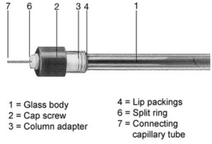
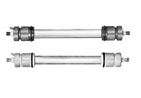
The name “Tsvet” means “colour” in Russian. In 1906, he coined the term “chromatography” from the Greek – combining chrōma (colour) and graphein (to write). The word is best translated loosely as “colour writing”. It has served as his lasting memorial ever since.
The Slow Rise of Column Chromatography
At first, though, Tsvet’s “chromatographic adsorption analysis”, as he called it himself, remained largely unknown, because he published exclusively in Russian. The mechanisms behind the separation were also far from understood. Around the same time, the German-Jewish biochemist Richard Willstätter (1872 – 1942) was investigating the structure of chlorophyll at the Kaiser Wilhelm Institute for Chemistry in Berlin. With paper chromatography, he succeeded in isolating the pigment and in determining its molecular formula. For this work on plant pigments, he received the Nobel Prize in Chemistry in 1915.
The column chromatography described by Tsvet only became more widely used in the 1930s. The German biochemists Edgar Lederer (1908 – 1988) and Richard Kuhn (1900–1967) developed it into a practical laboratory method in Heidelberg.
Chromatography Becomes a Standard Across Many Fields
The final breakthrough for chromatography came from understanding the separation mechanism. This insight grew out of the work of two British chemists in the 1940s: Archer John Porter Martin (1910 – 2002) and Richard Laurence Millington Synge (1914 – 1994). Drawing on experience from liquid extraction, they discovered how the separation actually works. Chromatographic separation depends on how differently substances distribute themselves between the stationary and the mobile phase.
It was soon used to decode the sequences of peptides. In doing so, it laid one of the cornerstones of modern biochemistry and molecular biology.
Size-Exclusion Chromatography
Size-exclusion chromatography separates molecules by size, as its name suggests. Depending on the solvent used, it is also known as gel-permeation chromatography or gel-filtration chromatography.
The analytes are separated by size as they pass through a porous gel. Small molecules enter the gel particles more easily. They therefore travel a longer path and elute later than larger ones. This makes the method particularly suitable for analysing plastics, where substances differ more by size than by their chemistry.
High-Performance Liquid Chromatography
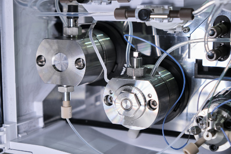
Biochemistry and natural-product chemistry deal with large molecules. For these, a further development of partition chromatography came into play: high-performance liquid chromatography (HPLC). Here, the separation process is accelerated. Two factors are decisive. The first is the high pressure of up to 400 bar. Secondly the use of very small stationary-phase particles, which offer a large surface area.
The peripheral equipment required for HPLC must meet equally high standards of pressure resistance and chemical inertness. The tubing therefore has to be especially pressure-resistant. Suitable options include PEEK tubing as well as titanium and stainless steel capillaries. These are joined using capillary connectors, which allow connection to columns, detectors and other instruments.
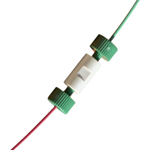
This high-pressure technique boosts the performance of column chromatography. Separation by hydrophobicity is not the only option, however. The method also helps to separate molecules by other properties, such as size, charge or affinity for a specific binding partner.
Thin-Layer Chromatography
Paper chromatography was later superseded by another powerful method: thin-layer chromatography. The technique dates back to 1938 and the work of two largely unknown Russians, N. A. Izmailov and M. S. Shraiber. In the early 1960s, the German chemist and pharmacist Egon Peter Gustav Stahl (1924 – 1986) refined it further. Working with the companies Merck (Darmstadt) and Degussa (Heidelberg), he established it as a near-universal analytical method for non-volatile substances. He also developed the technical equipment it required.
Thin-layer chromatography is technically simple and quick to carry out. Instead of paper, silica gels are the preferred stationary phase, along with aluminium oxides and modified celluloses. The stationary phase is applied in a thin layer to a flat support, usually a glass plate. An inert adhesive and binder, such as gypsum, holds it in place. The layer is then dried.
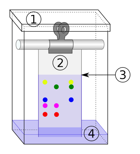
As in paper chromatography, the samples are applied to the dry, solid phase. The plate is then placed upright in a closed tank, with the solvent level well below the application point. Capillary forces draw the eluent up into the solid phase. This separates the substances according to how differently they adsorb to and desorb from that phase.
Gas Chromatography
In the late 1940s, the German physical chemist Erika Cremer (1900 – 1996) built on partition chromatography with her doctoral student Fritz Prior (1921 – 1996). Instead of a liquid, they used gases such as helium or nitrogen as the mobile phase. For the stationary phase, they used a high-boiling, viscous liquid on a porous, inert support – for example, silicone oils on silica gel. Their fundamental work led to a new type of column separation method: gas chromatography. It works for gaseous substances and for those that can be vaporised without decomposing.
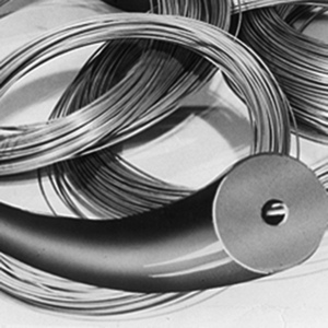
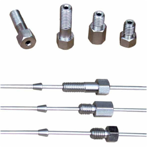
Its development into a powerful laboratory technique is closely linked to two British chemists: Archer John Porter Martin (1910 – 2002) and Anthony Trafford James (1922 – 2006). The 1950s also brought compatible detectors. These included the thermal conductivity detector, the flame ionisation detector, the electron capture detector and, finally, mass-spectrometric detectors. Together, they were decisive in establishing gas chromatography as a standard analytical and micro-preparative method.
Ion-Exchange Chromatography
Ion-exchange chromatography, also called ion chromatography, separates charged molecules, including nucleic acids and proteins. It does so according to their net charge, which depends on their isoelectric point. Their charge determines whether anion-exchange or cation-exchange chromatography is used. The principle is straightforward. The molecules bind ionically to an oppositely charged stationary phase. They are then eluted again by counter-ions from the eluent, at a suitable pH or ionic strength. An ion exchange therefore takes place.
In anion-exchange chromatography, negatively charged molecules – such as nucleic acids or proteins – bind to a positively charged stationary phase, for example a diethylaminoethyl (DEAE) resin. In cation-exchange chromatography, positively charged ions such as metal cations bind to a negatively charged stationary phase. One of the first applications came in the 1940s: the separation of rare-earth metals. These are hard to tell apart because of their chemical similarity. Ion-exchange chromatography is still used today to obtain pure rare-earth metals. These are indispensable raw materials for the entire field of microelectronics.
Affinity Chromatography
Affinity chromatography is widely used in the life sciences. It was developed in the 1960s by the Spanish-American pharmacologist Pedro Cuatrecasas (born 1935) and the Israeli biochemist Meir Wilchek (1936 – 2025). The method relies on the interactions between biomolecules and their ligands – for example, between an enzyme and its substrate, or between an antibody and an antigen. The biomolecule binds to its immobilised ligand. It is then eluted by deliberately changing conditions such as pH, salt concentration or the addition of competing ligands.
The method is also often used to purify genetically tagged proteins. These proteins carry a tag, such as a histidine tag or glutathione-S-transferase, that enables interaction with a specific solid phase. Affinity chromatography makes it possible to purify biomolecules very specifically, even in complex mixtures. It is also used to purify vaccines. Most of us have therefore benefited directly from the power of this technique at some point.
Ferdinand Runge’s Legacy Today
More than 150 years ago, Ferdinand Runge presented his chemical coats of arms and independently grown images to the public. He explained their formation through chemical interactions. Little could he have realised that he stood at the start of a dramatic development in the separation of substances.
Later generations of scientists took up his basic ideas and developed them further. Today, chromatography exists in many technically mature forms. It separates substances both on the micro scale, to analyse mixtures, and on the macro scale, to obtain pure substances. Without these modern chromatographic separation and purification methods, we would have to do without much that we value or even depend on. There would be no mobile phones, no specific vaccines and no specific medicines – and probably a great deal else besides.
Image sources:
Featured image | © sinhyu – stock.adobe.com
pH measurement | © Kim – stock.adobe.com
Schematic diagram of paper chromatography | © Theresa Knott, CC BY-SA 3.0 <https://creativecommons.org/licenses/by-sa/3.0>, via Wikimedia Commons
High-pressure tubing at the pump of an HPLC system | © vladim_ka – stock.adobe.com