 Reichelt Chemietechnik Magazine
« Sample Preparation in the Biochemical Laboratory »
The centrifuge is indispensable in any biochemical laboratory. Whether you work with cells, proteins or DNA, one moment always arrives: unwanted components need separating. Alongside other techniques such as filtration or chromatography, centrifugation is a well-established and easy method to apply.
How Centrifugation Works: The Role of Sedimentation
During centrifugation, solid particles are suspended in a liquid medium. A centrifugal force field then separates them by their different sedimentation rates. Sedimentation depends on the size, shape and density of each component. Larger particles settle faster than smaller ones. Likewise, denser particles settle faster than those of lower density. The medium itself also matters. Its density and viscosity influence how quickly particles sediment. In highly viscous media, particles settle more slowly than in thinner ones. A mixture of particles with varying sizes and densities can therefore be separated by spinning.

The Swedish chemist The (Theodor) Svedberg (1884–1971) won the Nobel Prize in Chemistry in 1926. He was the first to express these relationships mathematically. His formula, known as the Svedberg equation, could calculate the sedimentation rate of particles.
One value matters greatly in biochemistry: the sedimentation coefficient. It is the ratio of sedimentation rate to centrifugal acceleration. This quotient reflects a characteristic molecular property that can be measured experimentally. It is given in Svedberg units (S), where 1 S equals 10−13 seconds. The S-values of many biochemical particles are known. Some have even become part of a scientific name. One example is the 30S subunit of the prokaryotic ribosome.
Inside a Laboratory Centrifuge: Rotor, Tubes and Speed
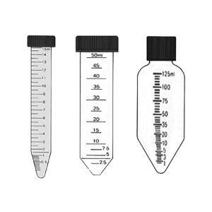
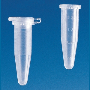
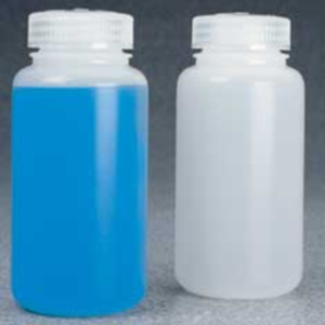
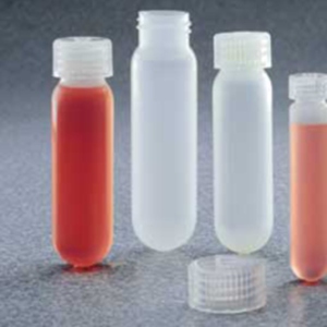
A centrifuge consists of a rotor driven by a motor to the required speed. The rotor holds the centrifuge vessels that contain the sample. These are either centrifuge tubes for small volumes or centrifuge bottles for larger amounts. Both are made from thick-walled glass or plastics such as PTFE, PP or HDPE. Their bases are either conical or rounded.
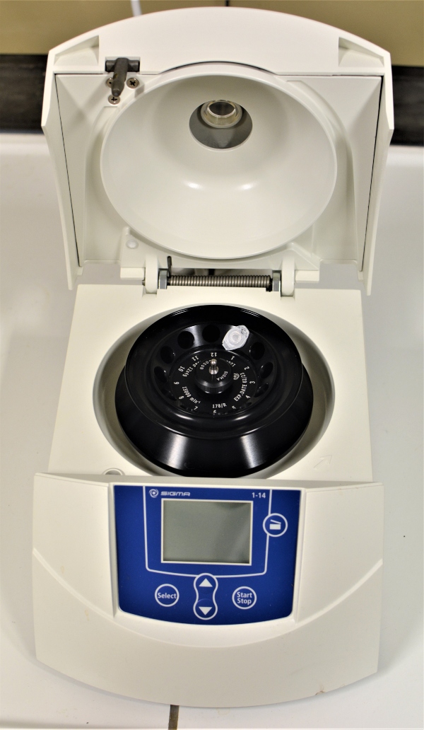
Rotor design depends on the sample volume and the intended use. Benchtop centrifuges separate samples in the microlitre and millilitre range. Larger centrifuges can hold sample volumes of up to several hundred millilitres.
The relative centrifugal acceleration g varies widely. It ranges from a few 103 g to several 106 g. Here, 1 g equals the acceleration of gravity. For very high speeds, ultracentrifuges are used. They run in a vacuum to avoid air resistance and the frictional heat it creates.
Centrifuge Rotor Types: Fixed-Angle, Vertical and Swing-Out
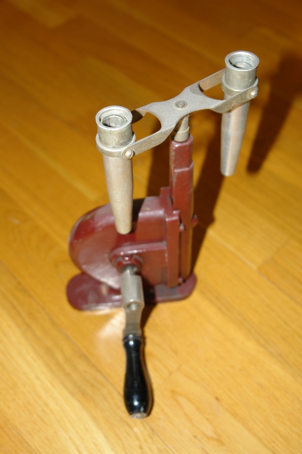
Many centrifuges use a fixed-angle rotor. Here the vessels spin at a set angle of about 30° to the rotor axis. In a vertical rotor, the sample tubes stand upright, perpendicular to the rotor axis. This type offers very short sedimentation paths and run times. It therefore suits applications with a high sample throughput.
One variant is the NVC rotor, where NVC stands for near vertical centrifugation. The vessels sit at a small angle to the rotor axis. Angles between 7.5° and 9° are common. This design is meant to make sample removal easier.
In a swing-out rotor, the vessels swing horizontally during the spin. They move outward, in the direction of the field. This rotor type is popular for separating large volumes.
Centrifugation Methods: Differential, Zonal and Isopycnic Techniques
Three techniques are used here: differential, isopycnic and zonal centrifugation. Each exploits a different physical parameter to achieve separation.
Differential Centrifugation: Separating Cell Fractions by Sedimentation Rate
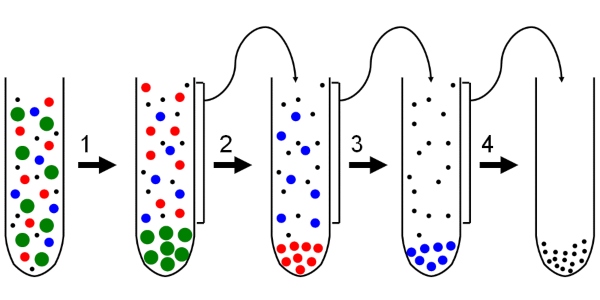
Differential centrifugation separates the components of a cell extract. It uses their different sedimentation rates. This method is always the first step in sample preparation. The sample is spun in stages at increasing g-values. Each supernatant then moves into the next centrifugation step. A typical scheme for separating a cell extract looks like this:
- At 1,000 g, whole cells and nuclei are spun down. They collect at the base of the vessel as a so-called pellet.
- The supernatant is then spun at 2,000 g. This pellets cell components such as mitochondria, peroxisomes and lysosomes.
- At 80,000 g, microsomes and small vesicles are separated from the rest. Large complexes such as ribosomes only sediment at 150,000 g, where they are collected as a pellet.
Differential centrifugation is therefore a very simple way to obtain different cell fractions.
Density Gradient Centrifugation: Zonal and Isopycnic Methods
Zonal centrifugation suits particles with similar sedimentation rates but different sizes. The sample is loaded onto a discontinuous density gradient, such as a sucrose gradient. This gradient consists of layers of rising concentration, typically between 18 and 30%. Their density increases accordingly. Carefully layering the solutions of different concentration creates the gradient. The densest sucrose solution sits at the base of the vessel.
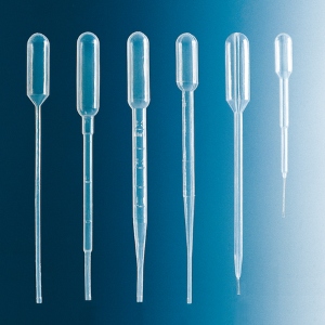
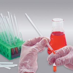
The sample is then spun at low speeds for a defined period. Because of the varying densities, the particles move at different sedimentation rates. They separate into discrete bands. Afterwards, these bands can be carefully drawn off with a Pasteur pipette.
It is important to stop the centrifugation after a set time. Otherwise, all particles would eventually travel to the bottom of the vessel.
Isopycnic centrifugation is also called sedimentation equilibrium centrifugation. It suits particles of similar size but different density. This technique also uses a density gradient. The gradient is not layered beforehand. Instead, it forms by itself as a continuous gradient during the spin. The run uses high g-values and takes time. A stable gradient sometimes forms only after 24 to 48 hours. Here the particles move through the gradient to one exact point. At that point, the density of the medium matches their own. In this so-called isopycnic zone, the sedimentation rate falls to zero. The particles therefore stop moving. As a result, the run time needs less careful attention than in zonal centrifugation.
In a continuous gradient, the particles in each layer are usually hard to see. They are therefore not drawn off from above with a pipette. Instead, a hole is pierced in the base of the vessel. The contents are then collected in fractions. A typical medium for a continuous gradient is a caesium chloride solution (CsCl). It can separate nucleic acids, for example.
Analytical Ultracentrifugation: Measuring Molecules and Complexes
The analytical ultracentrifuge is a less common instrument in the lab. It combines an ultracentrifuge with an optical detector. This is either a spectrophotometer or a refractometer. Instead of sample tubes, it uses UV-transparent cuvettes. Their windows are made of quartz or sapphire. The absorption of the solution is measured repeatedly, before and during the run. From this, the concentrations of the sedimenting particles can be determined. The readings can also reveal the exact sedimentation coefficients of molecules.
Another key area is the study of molecular complexes. Single molecules and those bound into complexes differ in their sedimentation rates.
This technique can study protein–protein or protein–DNA interactions, for example.
Alongside other methods of sample preparation and separation, centrifugation remains essential. It is still one of the most important and widely used techniques in the biochemical laboratory.